Saturday, February 27, 2010
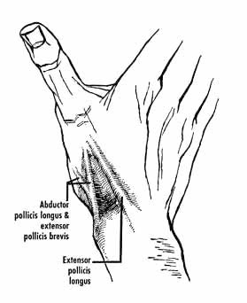
51 - Anatomical Snuff Box
*Anatomical snuff box is a triangular depression on the lateral aspect of wrist immediately distal to the radial styloid process, that becomes prominent when thumb is fully extended.
*The Contents of anatomical snuff box are :
- Cephalic vein
- Radial artery
- Superficial radial nerve
*Floor of the anatomical snuff box is formed by :
- Radial styloid
- Scaphoid (smooth convex articular surface)
- Trapezium
- Base of First metacarpal
*BOUNDARIES OF ANATOMICAL SNUFF BOX :
- Lateral/Anterior wall
~Abductor Pollicis Longus (Radially)
~Extensor Pollicis Brevis (Medially)
- Medial/Posterior wall
~Extensor Pollicis Longus
Tuesday, February 23, 2010
50 - Medial and Lateral Collateral Ligaments of Ankle
1. MEDIAL COLLATERAL LIGAMENT (OR DELTOID LIGAMENT) OF ANKLE :
*It consists of two sets of fibers, superficial and deep. Both parts have a common attachment above to the apex and margins of the medial malleolus. The lower attachment is indicated by the name of the fibers.
*Superficial fibers :
- The most anterior (tibionavicular) fibers pass forward to be inserted into the tuberosity of the navicular bone, and immediately behind this they blend with the medial margin of the plantar calcaneonavicular ligament (spring ligament).
- The middle (tibiocalcaneal) fibers descend almost perpendicularly to be inserted into the whole length of the sustentaculum tali of the calcaneum
- The posterior fibers (posterior tibiotalar) pass backward and laterally to be attached to the medial side of the talus, and its medial tubercle.
*Deep fibers :
- The deep fibers (anterior tibiotalar) are attached to the anterior part of medial surface of the talus.
*The deltoid ligament is crossed by the tendons of the tibialis posterior and Flexor digitorum longus.
2. LATERAL COLLATERAL LIGAMENT OF THE ANKLE :
*The lateral collateral ligament has 3 discrete bands or parts:
a. The anterior talofibular ligament - extends anteromedially from the anterior margin of the fibular malleolus to the neck of the talus.
b. The posterior talofibular ligament - extends almost horizontally from the lateral malleolar fossa to the lateral tubercle of the talus.
c. The calcaneofibular ligament - is a long cord which passes from a depression anterior to the apex of the fibular malleolus to a tubercle on the lateral calcaneal surface. It is crossed by the tendons of the peroneus longus and brevis.
----------------------------------------
Read this question from the May 2009 AIIMS Paper :
1q: Deltoid ligament is attached to all except :
a. Medial malleolus
b. Medial cuneiform
c. Spring ligament
d. Sustentaculum tali
-------------------------------
Saturday, February 20, 2010
49 - Medial and Lateral Menisci of Knee joint
*The menisci of the knee joint are two pads of cartilaginous tissue which serve to disperse friction in the knee joint between the lower leg (tibia) and the thigh (femur). They are shaped concave on the top and flat on the bottom, articulating with the tibia. They are attached to the small depressions (fossae) between the condyles of the tibia (intercondyloid fossa), and towards the center they are unattached and their shape narrows to a thin shelf.
*Both are cartilaginous tissues that provide structural integrity to the knee when it undergoes tension and torsion. The menisci are also known as 'semi-lunar' cartilages — referring to their half-moon "C" shape — a term which has been largely dropped by the medical profession, but which led to the menisci being called knee 'cartilages' by the lay public.
*The menisci act to disperse the weight of the body and reduce friction during movement. Since the condyles of the femur and tibia meet at one point (which changes during flexion and extension), the menisci spread the load of the body's weight. This differs from sesamoid bones, which are made of osseous tissue and whose function primarily is to protect the nearby tendon and to increase its mechanical effect.
SUMMARY:
*Both the Medial and lateral menisci are outside the synovial cavity but within the joint cavity.
*The Medial meniscus is larger than the lateral meniscus.
*The Medial meniscus is C shaped where as the lateral mensicus is circular shaped.
*The Medial meniscus is directly attached to the medial collateral ligament, where as the Popliteal muscle interferes in between the attachment of lateral meniscus and lateral collateral ligament.
*The Medial collateral ligament is most commonly injured when compared with the lateral collateral ligament.
Friday, February 5, 2010
Subscribe to:
Comments (Atom)


