The interpeduncular cistern (basal cistern or Fossa interpeduncularis) is a wide cavity where the arachnoid extends across between the two temporal lobes.
It encloses the cerebral peduncles and the structures contained in the interpeduncular fossa, and contains the arterial circle of Willis.
Diagram showing the positions of the three principal subarachnoid cisternæ. Interpeduncular cistern labeled at left center.
Tuesday, June 22, 2010
Monday, June 14, 2010
57 - Sulci and Gyri of Brain
*Lesion in the broca's area (inferior frontal gyrus) causes broca's aphasia (motor aphasia/expressive aphasia).
*Lesion in the wernicke's aphasia (supramarginal gyrus) of the parietal lobe and upper part of temporal lobe.
Friday, May 21, 2010
56 - AIIMS May 2010 Anatomy Mcqs
1. Middle superior alveolar nerve is a branch of
a) Mandibular division of trigeminal nerve
b) Palatine division of maxillary nerve
c) Anterior nasal division of maxillary nerve
d) Inferior alveolar nerve
2. All the following muscles retracts the scapula EXCEPT
a) Trapezius
b) Rhomboid major
c) Rhomboid minor
d) Levator scapulae
3. Cranial nerve NOT carrying parasympathetic fibres
a) 4th
b) 7th
c) 3rd
d) 9th
4. Prostatic urethra – True are A/E
a) Trapezoid in cross section
b) Elevated round swelling called verumontanum
c) Opening of prostatic ducts
d) Posterior part has urethral crest
5. Morgagni hernia presents most commonly on
a) Left posterior
b) Right anterior
c) Right posterior
d) Left anterior
6. Meralgia parasthetica is due to involvement of
a) Sural nerve
b) Medial cutaneous nerve of thigh
c) Lateral cutaneous nerve of thigh
d) Peroneal nerve
7. Celiac plexus is located
a) Anterolateral & around the aorta
b) Posterolateral & around the aorta
c) Anteromedial to lumbar sympathetic chain
d) Posterolateral to lumbar sympathetic chain
8. Paneth cells – True is
a) Rich in rough endoplasmic reticulum
b) High zinc content
c) Foamy cytoplasm
d) Numerous lysozyme granules
9. About sternocleidomastoid tumor all are true except –
a) Always associated with breech
b) Spontaenous resolution in most cases
c) Two-third have palpable neck mass at birth
d) Uncorrected cases develops plagiocephaly
10. The function of 8th cranial nerve is related to
a) Smell
b) Taste
c) Touch
d) Balance
11. Anterior ethmoidal nerve supplies all except?
a. Maxillary sinus
a) Interior of nasal cavity
b) Dural sheath of anterior cranial fossa
c) Ethmoidal air cells
12. A healthy young athlete is sitting at the edge of the table with knee at 90 degree
flexion. He fully extends it. What will happen?
a) Movement of tibial tuberosity towards lateral border of patella
b) Movement of tibial tuberosity towards medial border of patella
c) Movement of tibial tuberosity towards centre of patella
d) No change in position
13. Pain insensitive structure in brain is
a) Falx cerebri
b) Dural sheath surrounding vascular sinuses
c) Choroid plexus
d) Middle meningeal artery
14. Appendices epiploiceae present in
a) Appendix
b) Cecum
c) Rectum
d) Sigmoid colon
15. Pelvic splanchnic nerve supplies A/E
a) Appendix
b) Rectum
c) Uterus
d) Urinary bladder
Tuesday, May 18, 2010
55 - Pain Insensitive and Pain Sensitive structures in Brain
A. Pain Insensitive structures in Brain : (Intracranial)
1. Brain parenchyma
2. Ependyma
3. Choroid plexus
4. Piamatter
5. Arachnoid
6. Dura over convexity of skull ( Dura around vascular sinuses and vessels is sensitive to pain)
B. Pain Sensitive structures in Brain :
B1. Intracranial :
1. Cranial venous sinuses with afferent veins
2. Arteries at base of brain and arteries of dura including middle meningeal artery
3. Dura around venous sinuses and vessels
4. Falx cerebri
B2. Extracranial :
1. Skin
2. Scalp appendages
3. Periosteum
4. Muscles
5. Arteries
6. Mucosa
B3. Nerves :
1. Trigeminal (Fifth cranial nerve)
2. Facial (seventh cranial nerve)
3. Vagal (Tenth cranial nerve)
4. Glossopharyngeal (Ninth cranial nerve)
5. Second and Third cranial nerves
Monday, May 17, 2010
54 - Peroneus brevis
The peroneus brevis muscle (or fibularis brevis) lies under cover of the peroneus longus, and is a shorter and smaller muscle.
It arises from the lower two-thirds of the lateral surface of the body of the fibula; medial to the Peroneus longus; and from the intermuscular septa separating it from the adjacent muscles on the front and back of the leg.
The fibers pass vertically downward, and end in a tendon which runs behind the lateral malleolus along with but in front of that of the preceding muscle, the two tendons being enclosed in the same compartment, and lubricated by a common mucous sheath.
It then runs forward on the lateral side of the calcaneus, above the trochlear process and the tendon of the Peronæus longus, and is inserted into the tuberosity at the base of the fifth metatarsal bone, on its lateral side.
The terms "Peroneal" (i.e., Artery, Retinaculum) and "Peroneus" (i.e., Longus and Brevis) are derived from the Greek word Perone (pronounced Pair-uh-knee) meaning pin of a brooch or a buckle. In medical terminology, both terms refer to being of or relating to the fibula or to the outer portion of the leg.
Thursday, March 25, 2010
53 - Triangle of Auscultation
*The triangle of ausculation of the lungs is situated posterior and superficial to the scapula.
*It has the following boundaries:
- Superiorly, by the Trapezius
- Inferiorly, by the Latissimus dorsi
- Laterally by the medial margin of the scapula
*The floor is partly formed by the Rhomboideus major and parts of 6th and 7th ribs.
*The triangle of auscultation is a space on the back where the relatively thin musculature allows for respiratory sounds to be heard more clearly with a stethoscope.
*To better expose the floor of the triangle, which is made up of the posterior thoracic wall in the 6th intercostal space, the patient is asked to fold their arms across their chest, medially rotating the scapulae, while bending forward at the trunk.
Saturday, March 6, 2010
52 - Coronary arteries and Coronary veins
*The coronary arteries and the veins that drain into the coronary sinus. The posterior interventricular branch (PIV), although usually a branch of the right coronary artery (RC), may arise from the circumflex branch (C) of the left coronary artery (inset). In B, the left marginal vein can be seen ascending to join the great cardiac vein. The posterior vein of the left ventricle ascends and the oblique vein of the left atrium descends to end in the coronary sinus.
*AIV, anterior interventricular branch; C, circumflex branch; GC, great cardiac vein; LC, left coronary artery; MC, middle cardiac vein; PIV, posterior interventricular branch; Re, right coronary artery; S.-A, branch to sinuatrial node; SC, small cardiac vein.
Saturday, February 27, 2010
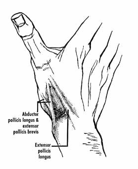
51 - Anatomical Snuff Box
*Anatomical snuff box is a triangular depression on the lateral aspect of wrist immediately distal to the radial styloid process, that becomes prominent when thumb is fully extended.
*The Contents of anatomical snuff box are :
- Cephalic vein
- Radial artery
- Superficial radial nerve
*Floor of the anatomical snuff box is formed by :
- Radial styloid
- Scaphoid (smooth convex articular surface)
- Trapezium
- Base of First metacarpal
*BOUNDARIES OF ANATOMICAL SNUFF BOX :
- Lateral/Anterior wall
~Abductor Pollicis Longus (Radially)
~Extensor Pollicis Brevis (Medially)
- Medial/Posterior wall
~Extensor Pollicis Longus
Tuesday, February 23, 2010
50 - Medial and Lateral Collateral Ligaments of Ankle
1. MEDIAL COLLATERAL LIGAMENT (OR DELTOID LIGAMENT) OF ANKLE :
*It consists of two sets of fibers, superficial and deep. Both parts have a common attachment above to the apex and margins of the medial malleolus. The lower attachment is indicated by the name of the fibers.
*Superficial fibers :
- The most anterior (tibionavicular) fibers pass forward to be inserted into the tuberosity of the navicular bone, and immediately behind this they blend with the medial margin of the plantar calcaneonavicular ligament (spring ligament).
- The middle (tibiocalcaneal) fibers descend almost perpendicularly to be inserted into the whole length of the sustentaculum tali of the calcaneum
- The posterior fibers (posterior tibiotalar) pass backward and laterally to be attached to the medial side of the talus, and its medial tubercle.
*Deep fibers :
- The deep fibers (anterior tibiotalar) are attached to the anterior part of medial surface of the talus.
*The deltoid ligament is crossed by the tendons of the tibialis posterior and Flexor digitorum longus.
2. LATERAL COLLATERAL LIGAMENT OF THE ANKLE :
*The lateral collateral ligament has 3 discrete bands or parts:
a. The anterior talofibular ligament - extends anteromedially from the anterior margin of the fibular malleolus to the neck of the talus.
b. The posterior talofibular ligament - extends almost horizontally from the lateral malleolar fossa to the lateral tubercle of the talus.
c. The calcaneofibular ligament - is a long cord which passes from a depression anterior to the apex of the fibular malleolus to a tubercle on the lateral calcaneal surface. It is crossed by the tendons of the peroneus longus and brevis.
----------------------------------------
Read this question from the May 2009 AIIMS Paper :
1q: Deltoid ligament is attached to all except :
a. Medial malleolus
b. Medial cuneiform
c. Spring ligament
d. Sustentaculum tali
-------------------------------
Saturday, February 20, 2010
49 - Medial and Lateral Menisci of Knee joint
*The menisci of the knee joint are two pads of cartilaginous tissue which serve to disperse friction in the knee joint between the lower leg (tibia) and the thigh (femur). They are shaped concave on the top and flat on the bottom, articulating with the tibia. They are attached to the small depressions (fossae) between the condyles of the tibia (intercondyloid fossa), and towards the center they are unattached and their shape narrows to a thin shelf.
*Both are cartilaginous tissues that provide structural integrity to the knee when it undergoes tension and torsion. The menisci are also known as 'semi-lunar' cartilages — referring to their half-moon "C" shape — a term which has been largely dropped by the medical profession, but which led to the menisci being called knee 'cartilages' by the lay public.
*The menisci act to disperse the weight of the body and reduce friction during movement. Since the condyles of the femur and tibia meet at one point (which changes during flexion and extension), the menisci spread the load of the body's weight. This differs from sesamoid bones, which are made of osseous tissue and whose function primarily is to protect the nearby tendon and to increase its mechanical effect.
SUMMARY:
*Both the Medial and lateral menisci are outside the synovial cavity but within the joint cavity.
*The Medial meniscus is larger than the lateral meniscus.
*The Medial meniscus is C shaped where as the lateral mensicus is circular shaped.
*The Medial meniscus is directly attached to the medial collateral ligament, where as the Popliteal muscle interferes in between the attachment of lateral meniscus and lateral collateral ligament.
*The Medial collateral ligament is most commonly injured when compared with the lateral collateral ligament.
Friday, February 5, 2010
Thursday, January 7, 2010
47 - Abdominal aorta mcqs with answers
1q: Ovarian artery arises from
a. Anterior part of abdominal aorta
b. Lateral part of abdominal aorta
c. Posterior part of abdominal aorta
d. None
2. All are lateral branches of abdominal aorta, except ?
a. Right testicular artery
b. Left renal artery
c. Inferior mesentric artery
d. Middle suprarenal artery
3. Cystic artery arises from ?
a. Right hepatic artery
b. Left hepatic artery
c. Common hepatic artery
d. Gastroduodenal artery
e. Inferior pancreaticoduodenal artery
4. The branches of the anterior division of internal iliac artery include :
a. Posterior gluteal artery
b. Uterine artery
c. Obturator artery
d. Internal pudendal artery
e. Iliolumbar artery
5. The gastroduodenal artery is derived from ?
a. Celiac artery
b. Hepatic artery
c. Splenic artery
d. Cystic artery
6. Watershed area between Superior mesentric artery and Inferior mesentric artery which commonly result in ishcemia is ?
a. Hepatic flexure
b. splenic flexure
c. Rectosigmoid junction
d. Ileocolic junction
Subscribe to:
Posts (Atom)


